 Zynfuse
A cranial defect is more than an opening in bone. Its location, shape, size, and surrounding tissue all affect how a repair is planned. Titanium Mesh For Cranial Repair is one option clinicians may consider because the material can be shaped to match a defect. The right choice, however, depends on the patient and the surgical plan—not on the material name alone. Small details matter.
This guide explains the practical factors behind mesh selection, including defect measurements, contour, mesh thickness, pore structure, and fixation options. Imaging helps the surgical team assess the area, while careful shaping can help the mesh follow the skull’s curves. The fit should be checked against the planned repair. Even a well-made implant may need adjustment; real anatomy rarely matches a perfect drawing.
The article also outlines questions to discuss with a qualified neurosurgeon, such as whether standard or patient-specific mesh is appropriate and what risks or follow-up may apply. Implant quality, manufacturing controls, and the team’s experience with the chosen system deserve attention. No mesh is right for every case. This overview offers general information, not a diagnosis or personal treatment recommendation. Individual decisions should be based on clinical evaluation and discussion with the treating team.
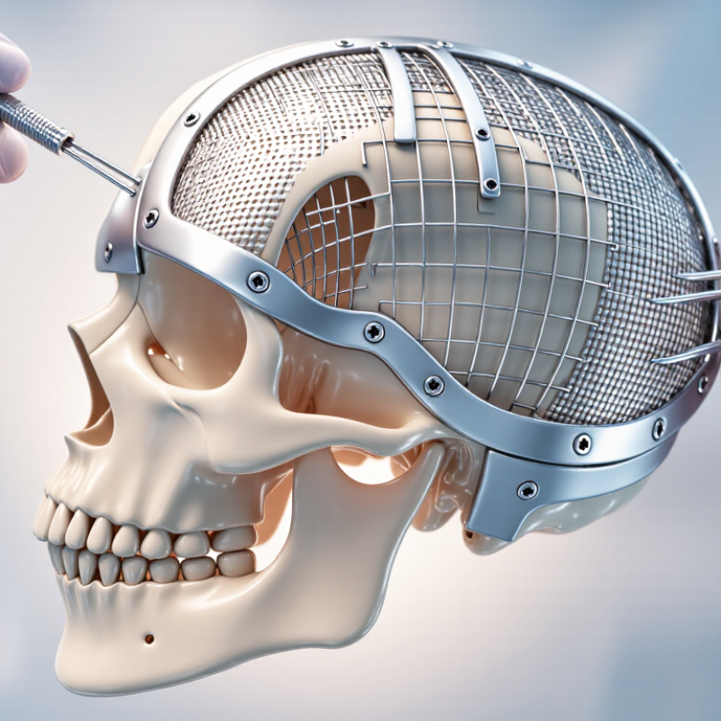
Titanium mesh provides a framework for rebuilding the skull’s contour after a portion of bone is missing. Its openings allow tissue to interact with the underlying surface, while the mesh can be shaped to follow the curve of the head. It is not bone. It does not restore brain tissue or replace the need for careful surgical planning. The goal is stable coverage and a contour that suits the defect and surrounding anatomy.
Mesh choice depends on the size and location of the opening, nearby nerves and vessels, skin condition, and the patient’s medical history. A thin, broad defect may call for different support than a small opening near the forehead. Surgeons also consider how the mesh will be fixed, whether its edges can be covered well, and how it may appear on imaging. Fit matters. A plan can look tidy on a scan, yet be less simple when the mesh meets real bone edges in surgery.
No single mesh design is right for every person. Porosity, thickness, stiffness, and contour all involve trade-offs; more rigid support is not automatically better. The treating neurosurgeon or craniofacial surgeon can explain why a specific option fits the defect, what complications are possible, and how healing will be monitored. Questions about pain, wound changes, or imaging should be discussed with the clinical team, since individual risks vary.
| Selection Dimension | What to Assess | Why It Matters | Practical Guidance |
|---|---|---|---|
| Defect size and location | Defect dimensions, skull curvature, nearby anatomy, and whether the repair is primary or revision surgery. | These factors affect contouring, coverage, fixation planning, and the choice between a standard or patient-specific reconstruction. | Use clinical examination and imaging to plan the reconstruction. The treating surgical team determines the appropriate design. |
| Mesh configuration | Flat, pre-contoured, or patient-specific mesh; available shape, coverage, and permitted intraoperative modification. | A suitable contour can help restore the intended cranial profile and reduce the need for extensive shaping during surgery. | Confirm the device’s instructions for use, including shaping limits and whether the selected configuration is intended for the planned application. |
| Thickness and mechanical properties | Specified thickness, rigidity, edge characteristics, and resistance to deformation for the particular device. | Mechanical behavior influences handling, contour maintenance, fixation, and protection of underlying tissues. | There is no universal thickness that suits every defect. Compare only dimensions and performance information provided for the specific device. |
| Pore pattern and open area | Mesh opening pattern, open area, and compatibility with the intended surgical technique. | The design affects flexibility and handling. Openings may also influence access for fixation and the relationship between the mesh and surrounding tissue. | Do not assume that one pore size is best for every patient; follow device specifications and the surgeon’s assessment of the defect. |
| Material and device documentation | The stated titanium material or alloy, applicable regulatory documentation, and any material-specific precautions. | Material composition and manufacturing details are device-specific and should be verified rather than inferred from the term “titanium mesh.” | Review the product labeling and instructions for use. Do not treat different titanium devices as interchangeable without clinical and regulatory review. |
| Fixation system | Compatible screws or other fixation components, available fixation points, and the quality of the surrounding bone. | Secure fixation is needed to maintain the planned position. Bone quality and anatomy can limit available fixation options. | Check component compatibility and follow the device instructions. Fixation location and technique are determined by the surgeon. |
| Soft-tissue coverage and wound status | Scalp condition, tissue coverage, prior surgery, infection history, and the overall wound environment. | Adequate soft-tissue coverage is an important part of reconstructive planning and can affect the risk of wound complications or implant exposure. | Assess the soft tissues and infection status before choosing a reconstruction plan; involve the appropriate surgical specialists when indicated. |
| Imaging and MRI considerations | Expected imaging needs, potential image artifact, and the exact implant’s MRI safety information. | Metal implants can affect image quality. MRI conditions and safety status depend on the specific device and its labeling. | Check the implant documentation before MRI and tell imaging staff about the implant. Do not assume MRI safety from material name alone. |
| Patient-specific factors | Age, medical conditions, medication use, healing considerations, allergies, and patient preferences. | These factors may influence operative planning, follow-up, and the balance of potential benefits and risks. | Discuss relevant medical history and treatment goals with the neurosurgeon or craniofacial surgical team. |
Note: This table is general educational information, not a device recommendation. Final selection should be made by a qualified surgical team using patient-specific assessment and the device’s current instructions for use.
Start with thin-slice CT images. They show the defect’s size, curve, and edges. Nearby sinuses, the orbit, and muscle also affect placement.
Yes. Thin, scarred, or previously infected skin may raise wound-healing and exposure concerns. A custom shape cannot fix poor tissue coverage. Easy to overlook.
Not always. It may follow an irregular skull contour more closely, but the surgeon must also assess coverage and fixation. There is no perfect template.
Lighter, open mesh may be easier to shape around curves. Thicker mesh can resist bending but may create a noticeable edge under thin skin.
No. A design that works near the forehead may not fit a delicate orbital rim. Location matters.
Ask about infection, wound breakdown, fluid collection, exposure, and visible contour changes. A smooth edge matters. So does coverage.
Reviews reported about 19.5% complications across different cranioplasty cases and materials. That is not a titanium-mesh-specific estimate or a personal prediction.
Follow-up may assess the incision, skin coverage, tenderness, swelling, or drainage. Seek clinical advice for fever, worsening pain, or an opening wound.
No. A scan can show implant position, but it cannot replace checking the skin. Long-term evidence remains limited, so expectations should stay realistic.
Choosing Titanium Mesh For Cranial Repair involves balancing the size and location of the skull defect with the patient’s anatomy, clinical needs, and planned procedure. Surgeons consider the contours of the surrounding bone, the protection required, and whether the reconstruction should be shaped during surgery or prepared in advance. These factors help guide the choice of mesh pattern, thickness, and design, since different options can offer varying levels of flexibility, support, and adaptability.
A suitable mesh should fit the defect closely, allow secure fixation, and work with the surgical approach and available instruments. Planning also includes considering potential complications, the patient’s recovery, and the need for follow-up imaging or clinical reviews. Long-term performance depends on an appropriate design, careful placement, and ongoing assessment. Decisions should be individualized by the treating surgical team, taking into account patient-specific risks and the goals of reconstruction.
